
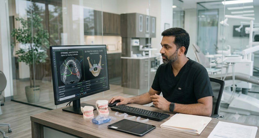
Introduction: The Quest for Sub-Millimeter Accuracy in Guided Surgery
The transition from freehand implantology to computer-assisted implant surgery (CAIS) represents one of the most substantial evolutionary leaps in contemporary surgical dentistry. By superimposing volumetric cone-beam computed tomography (CBCT) DICOM datasets onto optical surface impressions (STL/PLY), clinicians can engineer prosthetically driven implant sites before ever raising a flap.
However, clinical reality often challenges theoretical software models. Discrepancies between the virtual plan and the actual osteotomy bed—termed trajectory deviation—remain a critical concern, particularly in narrow interdental spaces, compromised ridge morphologies, and sites adjacent to vital neurovascular anatomy.
To master guided execution, clinicians must evaluate the biological and mechanical trade-offs between the two leading CAIS paradigms: Static Computer-Assisted Implant Surgery (s-CAIS) utilizing CAD/CAM 3D printed guides, and Dynamic Computer-Assisted Implant Surgery (d-CAIS) powered by real-time stereoscopic optical navigation.
1. The Tolerance Stack: Mechanical Variables in Static Guides
Static surgical guides provide a rigid, physically indexed template that constrains drill angulation and entry location via metal sleeves. While widely adopted and clinically predictable in routine scenarios, static guides remain vulnerable to tolerance stacking—the cumulative effect of micro-deviations introduced at multiple sequential stages.
The Cumulative Error Cascade
-
Imaging & Segmentation Artifacts: Patient motion artifacts, CBCT voxel resolution (e.g., 0.2mm vs. 0.4mm), and metal streak scattering distort tissue boundaries.
-
Surface Registration Errors: Minor mismatches during the alignment of the optical surface scan with the radiopaque markers or tooth structures in the DICOM dataset.
-
Additive Manufacturing Tolerances: Thermal shrinkage, resin contraction, and light-engine pixel calibration during 3D printing can create guide body distortion (typically 50–100 microns).
-
Mechanical Drill Play (The Key Culprit): To prevent friction and thermal osteonecrosis during osteotomies, a physical clearance gap must exist between the rotating drill shaft, the drill key/handle, and the guide sleeve. This gap typically ranges from 0.15mm to 0.25mm.
-
While this play seems negligible at the coronal entry point, angular displacement translates exponentially down the drill axis. At an implant apex 10–13mm subcrestally, an angular deviation of just 3° to 5° can translate into a linear apical shift of 1.2mm to 1.8mm.
-
-
Intraoperative Guide Instability: Tissue elasticity in mucosa-supported guides, micro-movement under surgical pressure, or incomplete seating in full-arch edentulous arches compound the mechanical error.
2. Dynamic Navigation Mechanics: Real-Time Optical Tracking
Dynamic navigation eliminates physical sleeves, templates, and 3D printing tolerances altogether. Instead of physically confining the handpiece, d-CAIS functions similarly to a real-time surgical GPS.
How d-CAIS Operates
-
Stereoscopic Optical Camera Array: An overhead multi-camera tracking unit emits overhead infrared or visible light tracking beams that read retro-reflective optical markers or active LEDs.
-
Patient Reference Tracker: A rigid tracking array is securely anchored to the patient’s dentition (via a thermoplastic bite stent) or directly to cortical bone via a mini-fixation screw in edentulous cases.
-
Handpiece Tracker: A secondary calibration array mounted directly to the surgical contra-angle handpiece tracks drill orientation, depth, and axis continuously at 30 to 60 frames per second.
-
Real-Time Visual Feedback: The surgeon watches a synchronized high-definition display showing cross-sectional, axial, and panoramic slices with a virtual crosshair targeting the pre-planned trajectory. The system gives instantaneous feedback on:
-
Entry point location
-
3D angulation alignment
-
Real-time drill depth (apical stop indicator)
-
Because there is no physical guide body obstructing the field, continuous external irrigation reaches the osteotomy site unimpeded, reducing the risk of bone overheating. Furthermore, if intraoperative bone quality reveals unexpected soft marrow spaces or buccal fenestrations, the trajectory can be modified in software without re-printing a surgical guide.
3. Anatomical Risk Management: Protecting Vital Structures in Deficient Ridges
When navigating anatomically unforgiving sites, the choice of guidance modality directly dictates clinical safety margins.
Managing the Inferior Alveolar Nerve (IAN)
In posterior mandibular atrophy where vertical bone height above the mandibular canal is limited (< 8mm), a standard apical deviation of 1.5mm in a static guide creates unacceptable neurosensory risks.
-
Dynamic Safety Buffers: Dynamic navigation enables custom audible and visual alerts when the drill tip approaches within 1.0mm of the mapped nerve canal. The clinician retains full peripheral tactile awareness while confirming safety across multiple anatomical planes.
Preventing Sinus Membrane Perforation & Maxillary Tilted Implants
In the atrophic posterior maxilla, engaging native palatal bone, the zygomatic buttress, or the pterygoid plates requires steep trajectory angles (25°–45°).
-
Physical static guides are often constrained by the patient’s maximum interincisal opening; long guided drills (often requiring 35–40mm of interarch clearance) cannot be inserted in limited-opening posterior sectors.
-
Dynamic navigation utilizes standard-length burs and open-access handpieces, enabling steep angulations and immediate apical anchorage without interocclusal space restrictions.
4. Clinical Comparison: Static Guides vs. Dynamic Navigation
| Clinical Parameter | Static 3D Printed Guides (s-CAIS) | Dynamic Surgical Navigation (d-CAIS) |
| Mechanical Drill Play | Inherent sleeve clearance (0.15–0.25mm) | Zero sleeve play (sensor tracking) |
| Interocclusal Clearance Required | High (demands guide height + sleeve + drill length) | Standard handpiece clearance |
| Intraoperative Flexibility | Rigid; plan cannot be modified chairside | Instantaneous on-screen plan recalibration |
| Direct Site Visualization | Partially obscured by guide body and sleeves | Unobstructed visual field and flap inspection |
| Irrigation Efficiency | Reduced due to guide bulk; requires careful flushing | Unrestricted external/internal saline delivery |
| Turnaround Time | Requires CAD design, printing, washing, and curing | Immediate surgery possible right after scan registration |
| Learning Curve | Low-to-moderate; tactile guidance | Moderate-to-high; indirect screen eye-hand coordination |
5. Clinical Takeaway: Selecting the Ideal Modality for Your Practice
Both static templates and dynamic navigation deliver substantially higher clinical accuracy and prosthetic passivity compared to freehand osteotomies. Selecting the right workflow depends on case anatomy and logistical requirements:
-
Choose Static 3D Guides When:
-
Performing routine single-tooth replacements or bounded short-span restorations where interocclusal opening is abundant.
-
Executing full-arch stackable guide cases (All-on-X) requiring sequential bone reduction, osteotomy delivery, and immediate provisional pickup indexed to bone pins.
-
You prefer a physical tactile stop without monitoring an external screen during drilling.
-
-
Choose Dynamic Navigation When:
-
Patients present with restricted mouth opening or posterior site limitations where long guided drills cannot clear opposing dentition.
-
The osteotomy runs within 1–2mm of high-risk boundaries (inferior alveolar nerve, mental loop, or deep sinus cavities).
-
Treating immediate extraction sockets where irregular extraction wall topography tends to deflect static drills off course.
-
Providing immediate, same-day surgical conversion where 3D printing and post-curing timelines are not feasible.
-
Elevate Your Surgical Workflows with Hanuka
At Hanuka, we support partner dental clinics with both end-to-end static guide fabrication and dynamic surgical planning workflows. From complex volumetric CBCT segmentations to chairside surgical alliance mentorship, our team ensures your high-value surgical cases achieve uncompromising prosthetic passivity and biological stability.
Connect with our surgical desk to evaluate your next guided surgery case.
